
Comprehensive Eye Exam
The Importance of the Comprehensive Eye Exam
A person may think they need an eye exam only when it’s time to update their eyewear prescription. The truth is, eye exams are about a lot more than seeing whether one needs a new pair of glasses or contacts. Comprehensive eye exams play an important role in overall wellness, and a person should get one annually for optimal vision health. Besides measuring vision, comprehensive eye exams can help identify early signs of certain chronic health conditions, including high blood pressure, diabetes, heart disease and high cholesterol. Many eye and vision problems have no obvious signs or symptoms, so a person might not know a problem exists. Early diagnosis and treatment of eye and vision problems can help prevent vision loss.
What is a Comprehensive Eye Exam?
A comprehensive eye exam can take approximately an hour and may vary slightly depending on the doctor and the complexity of tests required to fully evaluate a patient’s vision and the health of their eyes.
Here are the eye and vision tests that a patient is likely to encounter during a comprehensive eye exam:
Patient History
A patient will be asked about any current eye or vision problems and about overall health. The eye doctor will ask about medications, family medical history and any work-related or environmental conditions that may affect vision.
Visual Acuity
The Acuity test measures how clearly the patient can see. Each eye is tested separately. The patient will be asked to identify different letters of the alphabet on digital monitor positioned some distance away. The lines of type get smaller as one moves down the chart. The results of visual acuity testing are written as a fraction, such as 20/40. The top number in the fraction is the standard distance at which testing is
done (20 feet). The bottom number is the smallest letter size you were able to read. A person with 20/40 visual acuity would have to get within 20 feet to see a letter that should be seen clearly at 40 feet. Normal distance visual acuity is 20/20.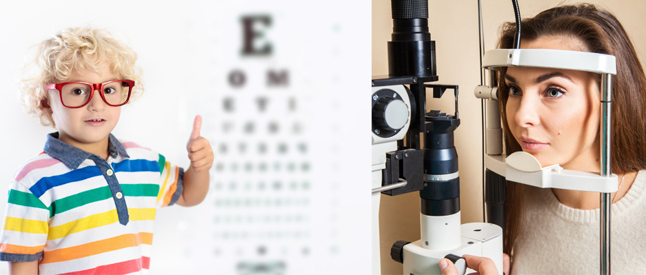
Visual Function Tests
Eye Movement/ Ocular Motility: The eye doctor evaluates the muscles that control eye movement by watching a patient’s eyes follow a moving object, such as a pen or small light. The doctor is looking for signs of muscle weakness, poor control or poor coordination. Problems with eye movements can cause eye strain and may affect reading ability and other activities such as sports.
Color Test: A screening test that checks for color blindness. In addition to detecting hereditary color vision deficiencies, color blind tests also alert the eye doctor to possible eye health conditions that may affect color vision.
Eye Alignment/ Cover Test. The patient is asked to focus on a target object across the room and cover each of eye alternately. During the test the eye doctor will assess whether the uncovered eye moves to pick up fixation on the target object. The cover test reveals whether the eyes work together and could indicate conditions such as strabismus (crossed eyes) or other problems that could cause eye strain or amblyopia (“lazy eye”).
Peripheral/Side Vision: Peripheral vision testing requires a patient to stare into a perimeter machine and react to a series of blinking lights in the outer visual field, or special cards with specific lines and patterns that create forced optical illusions. Since peripheral vision loss can be a sign of a number of eye diseases, including glaucoma and other optic nerve disorders, side vision must be tested regularly.
Pupil Reflex Test: The test involves the patient being asked to look at a target object across the room while the eye doctor shines a light into each eye. The doctor is watching for the pupils to constrict in response to the light, making note of the size and shape of the pupils.
Depth Perception/Stereopsis test: Stereopsis is the term used to describe eye teaming that enables normal depth perception and appreciation of the 3-dimensional nature of objects. The patient is asked
to wear a pair of “3D” glasses, look at a booklet of test patterns and identify the “closer” circle in each pattern. Eye teaming skills that should enable the patient to experience normal depth perception.
Slit-lamp examination: A slit lamp is a microscope that magnifies and illuminates the front of the patient’s eye. This device enables the eye doctor to examine the eyelids, lashes, cornea, iris, lens and fluid chambers between your cornea and iris. A wide range of eye conditions and diseases can be detected with the slit lamp exam, including cataracts, macular degeneration and diabetic retinopathy.
Refraction
A refraction test determines the lens power needed to compensate for any refractive error (nearsightedness, farsightedness or astigmatism) that may exist and the exact eyeglass prescription needed.
During a refraction, the eye doctor places an digital instrument called a phoropter in front of the patient’s eyes and a series of lens options will be presented. The patient will be asked to judge which of the two lenses provides clearer vision when viewing an eye chart. The refraction determines the patient’s level of hyperopia (farsightedness), myopia (nearsightedness), astigmatism and presbyopia.
We use digital Autorefractors to get us a remarkably close starting point to the final prescription. It only takes 2-3 minutes with the digital phoropters to produce crisp, clear and comfortable vision with new glasses or contact lenses. Digital instruments provide a faster, easier for the patient and more accurate refraction test.
Tonometry / Glaucoma Test
Glaucoma typically does not present any warning signs until the patient experiences significant vision loss. Tonometry measures the fluid pressure inside your eye (intraocular pressure). This is one test that helps the eye doctor detect glaucoma, a disease that damages the optic nerve.
Instead of the old ‘puff test’ our office uses the latest technology iCare tonometer that allows painless measurement the pressure in the patient’s eye without the need for drops and no air puff. There is no patient hesitation when the test is performed on the patients second eye. It provides accurate measurement of eye pressure of even the most demanding patients such as children or patients with dementia. Patients with high eye pressure, may be at risk for or have glaucoma.
Retinal Examination / Ophthalmoscopy / Retinal Imaging
This examination — sometimes called ophthalmoscopy or fundoscopy — allows the eye doctor to evaluate the entire back of the eye, including the macula, peripheral retina, the optic disk and the retinal blood vessels that nourish the retina. This test is sometimes done in conjunction with Pupil Dilation. Instead of pupil dilation, most patients prefer our iWellness retinal exam. In 0.50 seconds we capture a beautiful wide-field retinal image of each eye using our Optomap. Then in another 0.50 seconds our OCT imaging produces a spectacular cross-sectional view of the macula, the area of the retina for central vision. Visualization of the retina can provide lots of information about a medical diagnosis. Imaging helps educate our patients and allows us to better confirm early diagnoses of eye diseases such as glaucoma, macular degeneration, retinal tears or detachments as well as systemic diseases such as high blood pressure, diabetes, increased fluid pressure in the brain and infections like endocarditis.
Vision Source Insight Eyecare – Comprehensive Eye Exam
There is so much more to an eye exam than just updating an eyewear prescription. The eye health and broader medical benefits of an eye exam are considerable and can offer early diagnosis for a series of conditions. Be sure to schedule you appointment today. Vision Source Insight Eyecare provides comprehensive eye exams for Adults and Pediatrics patients in Sandy Springs, Dunwoody, Buckhead and Atlanta.